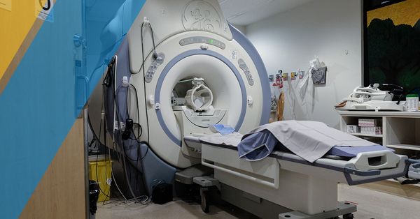
Magnetic Resonance Imaging (MRI) scans are a vital part of any radiologist’s tools for viewing, diagnosing, and treating any issues that patients may be suffering from. Last time, we looked at the uses of MRI imaging and how they can be preferable to CT scans. North Pittsburgh Imaging Specialists is your source for Pittsburgh MRI services, along with other imaging specialties. Today, we’ll look deeper into how MRI scans work and the science surrounding that big tube.
The Science Behind Your Pittsburgh MRI Scans Part 2
North Pittsburgh Imaging Specialists
How Does It Work?
Science. The longer answer is that MRI machines utilize two giant magnets that make up the most important part of the operation. Humans typically consist of around 60% moisture, and this abundance of water molecules comes into play with magnetic imaging. Normally, your molecules are scattered about randomly with no discernible pattern. Upon entering an MRI machine, one of the magnets will pull the hydrogen in your water and fat in one direction, either spinning towards your feet or head. At this point, the atoms will tend to cancel each other out if moving in opposite directions.
Once we have the atoms set in a northern or southern direction, we then focus on the area being scanned. The machine will send a radio frequency (RF) pulse toward the area in question. This RF pulse is specially tuned to interact with hydrogen. Once the pulse is activated, the protons in the selected area will receive the energy from that pulse and start to spin in the opposite direction. The new direction of travel for these target atoms is how derive the term “resonance”. The speed and direction in which the pulse sends these molecules on their journey is based on the specific part of the body. This frequency determination is known as the Larmor frequency, and is set based on the strength of the magnetic field that it is working in.
At this point, the other magnet (and its subsidiaries) activates in a series of pulses, activating each atom in the selected area and sending them temporarily in a different direction. The result is a change in movement, which helps to paint a magnetic image on the computer based on the consistency of the tissue. When the machine is turned off, your hydrogen protons will start to ease back into their natural states, no longer gravitating towards the head or feet. This movement produces a signal that is picked up by the machine and sent to the computer for compilation. Technology allows these signals to be combined to create a two or three-dimensional map of the topography of your tissues. The key part of this phase is the observation of differences between normal and abnormal body tissues. Contrast dyes will react differently based on the tissue type and can be observed on screen by the human eye.
The full amount of information behind MRI tests would be enough to fill thousands of blog pages. Our quick synopsis is here to inform and hopefully entertain those who are interested in magnetic resonance imaging. Next time, we’ll look at how MRIs work for patients and what to expect when you’re due for a scan. If you’re in need of a Pittsburgh MRI specialist, don’t hesitate to contact us today to set up an appointment!
Categories: